About the Journal
Scope of the Journal
Abstracting & Indexing
About the Journal
Archive
Author Instructions
Current issue
Editorial Board
JoMI Policy
JoMI Policy - A to C
JoMI Policy - D to L
JoMI Policy - O to T
Submit Manuscript
Updates
JoMI Updates 2026
JoMI Updates 2025
JoMI Updates 2027
Visit Us


CASE REPORT
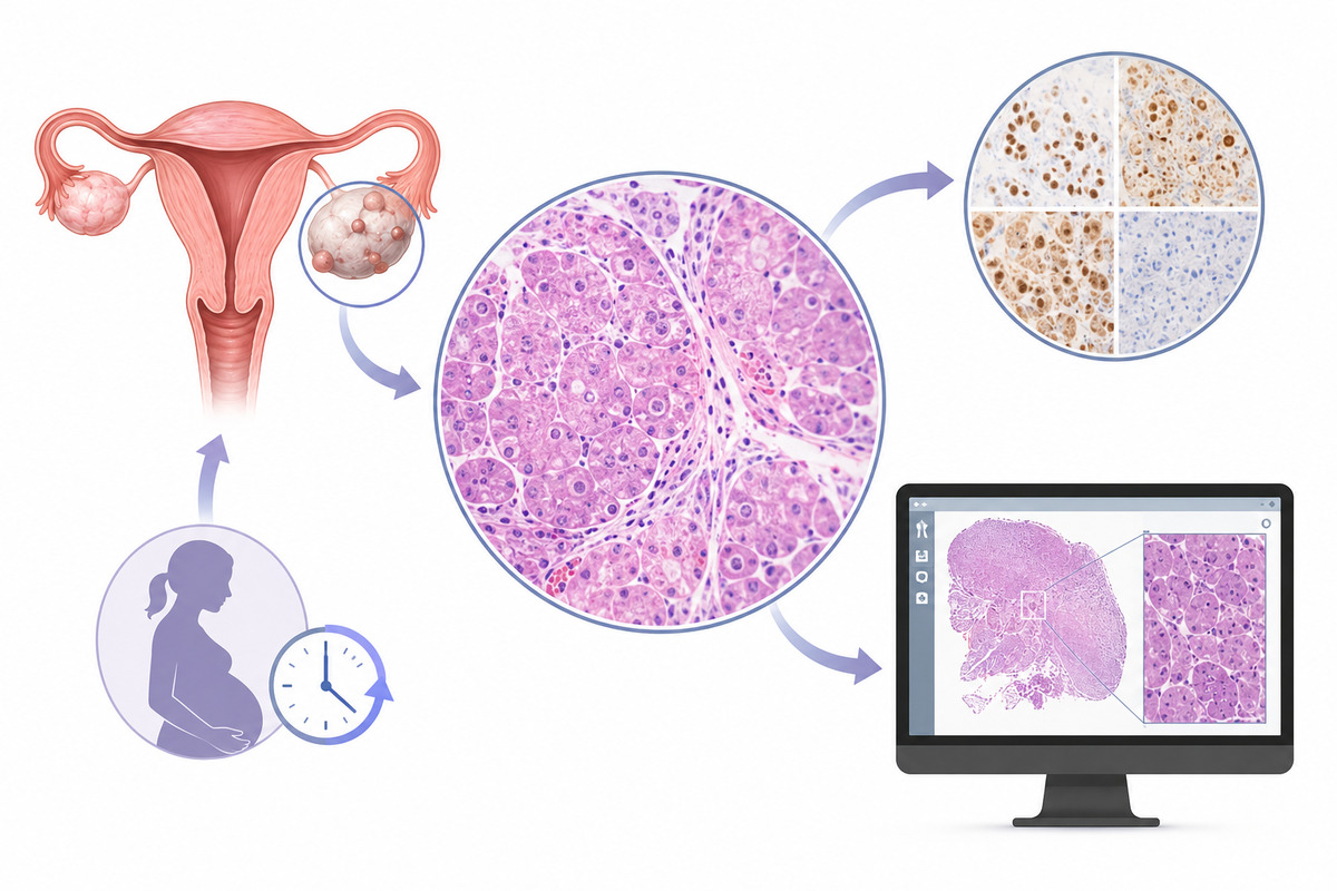
Ovarian Deciduosis Mimicking Malignancy: A Case Report & Digital Pathology Framework for Recognizing Benign Pregnancy-Associated Lesions
1
Pathology, All India Institute of Medical Sciences, Nagpur, India
2
Obstetrics & Gynecology, All India Institute of Medical Sciences, Nagpur, India
Submission date: 2026-05-10
Final revision date: 2026-06-21
Acceptance date: 2026-07-06
Online publication date: 2026-07-28
Publication date: 2026-07-28
Corresponding author
Journal of Medico Informatics 2026;02(Issue 03):4-8
HIGHLIGHTS
- Ovarian deciduosis can closely mimic disseminated malignancy during pregnancy.
- Histology revealed bland decidual cells without atypia or mitoses.
- Immunohistochemistry supported benign progesterone-driven decidual differentiation in ovarian tissue.
- Digital pathology may reduce diagnostic errors involving rare benign mimics.
- Structured reporting and registries could strengthen recognition of uncommon deciduosis.
KEYWORDS
computer-aided diagnosisdigital pathologyOvarian deciduosisectopic decidual reactionsynoptic structured reporting
TOPICS
ABSTRACT
Ectopic decidualization, or deciduosis, is a benign, hormone-dependent proliferation of decidual-type stromal cells outside the uterine endometrium. During pregnancy, it is most often an incidental finding. Yet, its nodular gross appearance and cellular histology can closely imitate disseminated malignancy, exposing patients to overtreatment when it is misread intra-operatively or on biopsy. A 34-year-old second-gravida woman was admitted in active labour and had an uncomplicated full-term vaginal delivery. On the second postpartum day, during elective tubal ligation, a 5 × 4.5 × 3 cm right ovarian cyst studded with off-white, shiny surface nodules was identified and excised together with bilateral tubal ligation. Histology demonstrated a serous cystadenoma lined by a single layer of flattened-to-cuboidal epithelium, accompanied by surface and parenchymal nodules of bland, large polygonal cells with abundant eosinophilic cytoplasm consistent with decidua. Immunohistochemistry (PR positive, CD10 positive, focal inhibin positivity, CK20 negative) together with the absence of nuclear atypia or mitoses supported a benign decidual reaction. A systematic search excluded occult malignancy; after multidisciplinary consensus, no further treatment was given, and the patient remained asymptomatic. We situate this case within a medico-informatics framework, proposing that whole-slide imaging with computer-aided image analysis can reduce the diagnostic-error risk posed by benign mimics while generating reusable, machine-readable evidence for rare entities. Clinician and pathologist awareness of the condition, stringent histological criteria, and digital diagnostic infrastructure together protect pregnant patients from unnecessary intervention.
ABBREVIATIONS
AI: Artificial Intelligence
CA-125: Cancer Antigen 125
CEA: Carcinoembryonic Antigen
CD10: Cluster of Differentiation 10
CK7: Cytokeratin 7
CK20: Cytokeratin 20
H&E: Haematoxylin and Eosin
IHC: Immunohistochemistry
ICD-O: International Classification of Diseases for Oncology
Ki-67: Ki-67 Proliferation Marker
PET-CT: Positron Emission Tomography–Computed Tomography
PR: Progesterone Receptor
SF-1: Steroidogenic Factor 1
SNOMED CT: Systematized Nomenclature of Medicine Clinical Terms
WSI: Whole-Slide Imaging
ACKNOWLEDGEMENTS
The authors sincerely thank the Department of Pathology and the Department of Obstetrics & Gynecology, All India Institute of Medical Sciences (AIIMS), Nagpur, for their support in the diagnosis, clinical management, and preparation of this case report. The authors also acknowledge the patient for providing informed consent for publication.
FUNDING
This research received no external funding. All work was conducted using institutional resources without dedicated grant support.
CONFLICT OF INTEREST
The authors declare that they have no known financial, personal, academic, or other relationships that could inappropriately influence, or be perceived to influence, the work reported in this manuscript. The authors confirm that there are no competing interests to declare.
PEER REVIEW INFORMATION
Article has been screened for originality
© 2026 The Authors. Published by Aayvu Publications Private Limited under CC BY 4.0.
REFERENCES (10)
1.
Bellizzi AM. (2020), An Algorithmic Immunohistochemical Approach to Define Tumor Type and Assign Site of Origin, Adv Anat Pathol, 27(3):114-163. doi:10.1097/PAP.0000000000000256. PMID: 32205473.
2.
Eloy C, Seegers P, Bazyleva E, Fraggetta F. (2024), The 1 million words pathology report or the challenge of a reproducible and meaningful message, ESMO Real World Data Digit Oncol, 4:100044. doi:10.1016/j.esmorw.2024.100044. PMID: 41647787.
3.
Kaneko M, Nozawa H, Rokutan H, Murono K, Ushiku T, Ishihara S. (2021), Ectopic decidua of the appendix: a case report, Surg Case Rep, 7(1):117. doi:10.1186/s40792-021-01204-9. PMID: 33973073.
4.
Kennedy NT, Sebastian A, Thomas DS, Thomas A, Gupta M, Kumar RM, Peedicayil A. (2019), Diagnostic Accuracy of Frozen Section and Its Influence on Intraoperative Management of Indeterminate Epithelial Ovarian Tumors, Indian J Surg Oncol, 10(2):268-273. doi:10.1007/s13193-018-00869-3. PMID: 31168246.
5.
Kinra P, Sen A, Sharma JC. (2006), Ectopic Decidual Reaction: A Case Report, Med J Armed Forces India, 62(3):280-1. doi:10.1016/S0377-1237(06)80022-5. PMID: 27407910.
6.
Mangla M, Nautiyal R, Shirazi N, Pati B. (2021), Ectopic Cervical Deciduosis: A Rare Cause of Antepartum Hemorrhage in Mid Trimester, Eurasian J Med, 53(2):152-154. doi:10.5152/eurasianjmed.2021.20163. PMID: 34177301.
7.
Masjoodi S, Anbardar MH, Shokripour M, Omidifar N. (2025), Whole Slide Imaging (WSI) in Pathology: Emerging Trends and Future Applications in Clinical Diagnostics, Medical Education, and Pathology, Iran J Pathol, 20(3):257-265. doi:10.30699/ijp.2025.2044210.3367. PMID: 40746923.
8.
McCluggage WG. (2006), My approach to the interpretation of endometrial biopsies and curettings, J Clin Pathol, 59(8):801-12. doi:10.1136/jcp.2005.029702. PMID: 16873562.
9.
Selak N, Cerkez I, Iljazovic E, Sadikovic A, Konrad Custovic M, Mustedanagic Mujanovic J, Ahmetovic Karic E. (2025), Extraovarian fibrothecomas: Two case reports and comprehensive review of ovarian sex cord-stromal fibroma-thecoma tumors, Biomol Biomed, 26(3):509-524. doi:10.17305/bb.2025.12816. PMID: 40828576.
10.
Sorokin P, Nikiforchin A, Panin A, Zhukov A, Gushchin V, Kurtser M. (2020), Diffuse Ectopic Deciduosis Imitating Peritoneal Carcinomatosis with Acute Abdomen Presentation: A Case Report and Literature Review, Case Rep Obstet Gynecol, 2020:8847082. doi:10.1155/2020/8847082. PMID: 33062356.
| ISSN: | 3108-2696 |

We process personal data collected when visiting the website. The function of obtaining information about users and their behavior is carried out by voluntarily entered information in forms and saving cookies in end devices. Data, including cookies, are used to provide services, improve the user experience and to analyze the traffic in accordance with the Privacy policy. Data are also collected and processed by Google Analytics tool (more).
You can change cookies settings in your browser. Restricted use of cookies in the browser configuration may affect some functionalities of the website.
You can change cookies settings in your browser. Restricted use of cookies in the browser configuration may affect some functionalities of the website.